
Radiotherapy is one of the most important anti-cancer treatments and is used to treat around half of all patients in Europe. In radiotherapy, beams of ionising radiation are used to target and kill cancerous cells with a high radiation dose. The more precise this can be done, the higher the survival rates and lower the undesired site-effects.
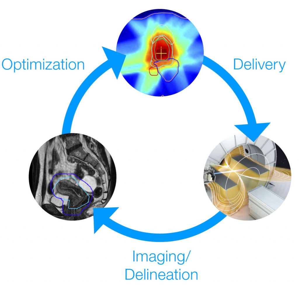
Precision is limited if accurate and up-to-date imaging of the targeted tumor is lacking. Combining real-time Magnetic Resonance (MR) imaging with radiotherapy (Magnetic resonance guided radiotherapy – MRgRT), provides more detailed images and anatomical information of a patient during treatment than conventional techniques. The simultaneous use of MR images and irradiation allows for more precise dose delivery and continuous optimisation of the dose distribution. Even organ or tumour motion, caused by internal movements of the patient (e.g. breathing, swallowing), during treatment can potentially be corrected for, increasing the accuracy of the targeting. This new technique also reduces the side-effects of the treatment and avoids additional exposure to harmful radiation from diagnostic imaging modalities (e.g. CT) currently in use.
Currently the number of clinical MR-guided X-ray Therapy (MRgXT) facilities (combining x-ray beams and MRI) is increasing rapidly. Although not as advanced clinically, future developments are also being made with MR-guided Proton Therapy (MRgPT) which combines proton beams and MRI.
Accurate measurements of the radiation dose (dosimetry) is a prerequisite for safe and effective radiotherapy treatments. For modern radiotherapy techniques, small radiation fields (field size < 3 cm) are extensively used. The recent Code of Practice (CoP) TRS-483, enables medical physicists to perform traceable small field dosimetry for these techniques. In MRgRT, dosimetry needs to be performed in the presence of the magnetic field of the MRI scanner, which is known to affect both the dose distribution and the calibration of detectors used for dosimetry. For MRgRT, small radiation fields are equally important, however existing CoPs for small field dosimetry are inadequate for application in MRgRT.
MRgRT II
In this second MRgRT project we will develop a methodology for small field dosimetry in MRgXT facilities with an uncertainty similar to that for conventional radiotherapy (2.0 %). The project also includes a method for traceable dose measurement in MRgPT to show the feasibility of dose delivery with MRgPT and to prepare for (pre)-clinical investigations. These results will accelerate the development of Codes of Practice for small field dosimetry in MRgRT.
MRgRT I
The first MRgRT Joint Research Project focused on developing new standards and measurement methods for reference dosimetry (on 10 x 10 cm2 reference fields). The project has established metrological capabilities in reference dosimetry, imaging and quality assurance procedures for clinical MRgRT treatments. This is not only vital for the safe clinical implementation of MRgRT, but also for future innovations in MRgRT. Moreover, it has paved the way to perform reference dosimetry in MRgRT with a similar uncertainty as for conventional radiotherapy.